Fig. 4.
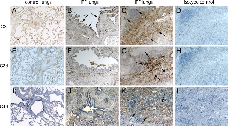
Immunohistochemistry of complement component 3 (C3), C3d, and C4d. Normal lungs are stained by C3, C3d, and C4d (A, E, and I). Idiopathic pulmonary fibrosis (IPF) lungs are stained by C3 (B and C), C3d (F and G), and C4d (J and K) (arrows). Isotype controls for C3, C3d, and C4d are shown (D, H, and L). Airway epithelial cells (B, F, and J) and interstitial lesions (C, G, and K) are shown. In IPF lung tissue C3 was expressed in airway epithelial cells, vascular walls, and interstitial lesions (B and C). C3d and C4d were expressed in the vascular walls and interstitial lesions but not in airway epithelial cells (F, G, J, and K). Brown color, DAB; blue color, hematoxylin. Magnification: ×4.

