Fig. 5.
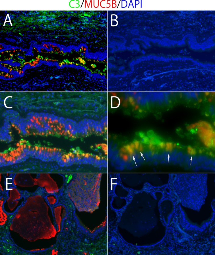
Immunofluorescence double staining of complement component 3 (C3) and MUC5B in human lung tissue. Double positive cells (arrows) denote airway epithelial cells (A, C, and D). There is no C3 expression in honeycomb cysts (E). B and F are isotype controls for A and E, respectively. Magnifications: A, B, E, and F: ×10; C: ×20; D: ×40. Fluorescent colors: green, C3 staining; red, MUC5B staining: yellow, overlay; blue, DAPI.

