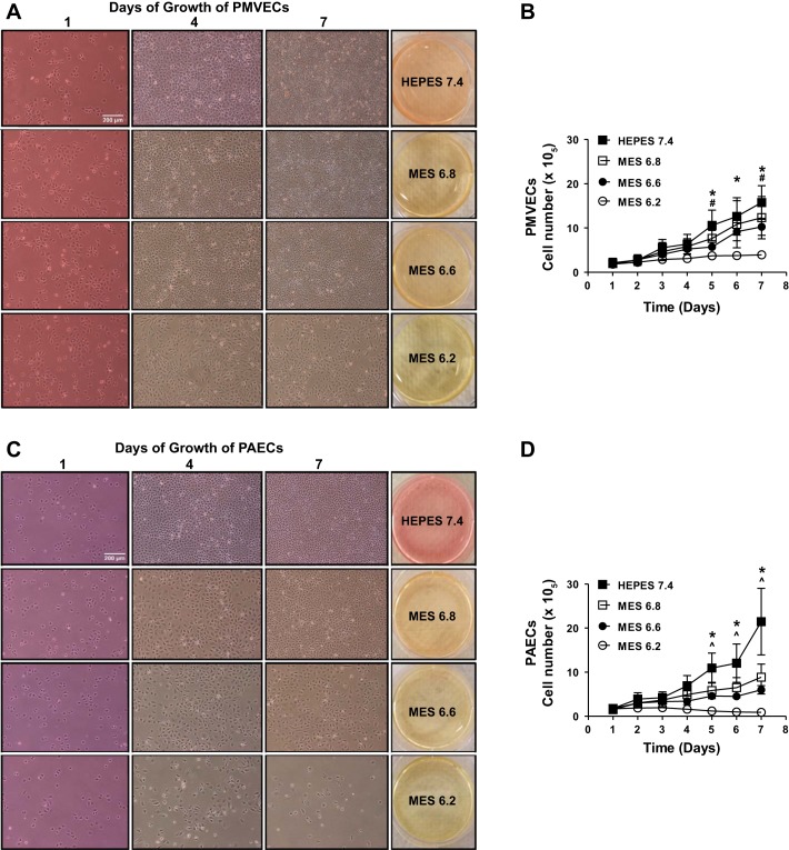
Fig. 1.
Pulmonary microvascular endothelial cells (PMVECs) survive better than do pulmonary arterial endothelial cells (PAECs) in acidosis. PMVECs and PAECs were seeded at 1.0 × 10⁵ cells/well on 6-well plates on bicarbonate-buffered media. 1 day after cell seeding, media was changed to either HEPES-buffered pH 7.4 or 2-(N-morpholino)ethanesulfonic acid (MES)-buffered pH 6.8, 6.6, or 6.2 media. Cells were imaged and counted with daily media change. A and B: PMVECs grew to confluence and stayed healthy for 7 days in pH 7.4, 6.8, and 6.6, but the cells underwent growth arrest in pH 6.2. C and D: PAECs grew to confluence and stayed healthy for 7 days in pH 7.4 but showed visible cell count decrease in pH 6.8, 6.6, and 6.2 in a dose-dependent manner. Data represent means ± SD. 2-way ANOVA and Bonferroni post hoc tests were used to compare between different pH groups. For each cell type and pH group, at least 5 separate experiments were performed. *Significant difference (P < 0.05) in 7.4 vs. 6.2 groups. #Significant difference (P < 0.05) in 7.4 vs. 6.6 groups. ^Significant difference (P < 0.05) in 7.4 vs. 6.8 groups.

