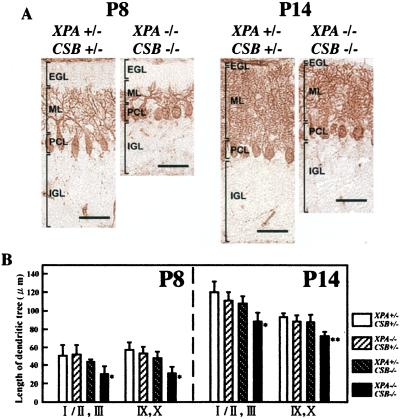
Figure 4.
Immunohistochemical staining of cerebellar PCs with anticalbindin antibody. (A) PC morphologies of XPA+/−CSB+/− and XPA−/−CSB−/− mice at P8 (Left) and P14 (Right). EGL, external granule cell layer; PCL, PC layer; IGL, internal granule cell layer; ML, molecular layer. (Bar = 50 μm.) (B) Measures of the length of the dendritic tree of PCs in lobules I/II and III, and IX/X of each genotypic mice cerebellum at P8 and P14. Values are mean ± SD for six midsagittal sections derived from five to eight mice of each genotype. *, P < 0.001, and **, P < 0.0001, against XPA+/−CSB+/−.

