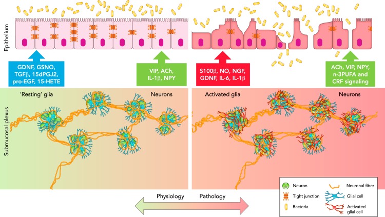
FIGURE 2.
Schematic representation of the neuronal-glial-epithelial unit in the GI tract
In physiological conditions (left), glial cells (blue) surround neurons (green) to form ganglia, which are interconnected through neuronal fibers (orange) to form the submucosal plexus. “Resting” glia communicate with epithelial cells via the release of GDNF, GSNO, TGFβ1, 15dPGJ2, pro-EGF, and 15-HETE to preserve barrier function, whereas submucosal neurons release VIP, ACh, IL-1β, and NPY. In pathological conditions (right), glial cells become activated (red) and release S100β, NO, NGF, GDNF, IL-6, and IL-1β, which alter epithelial barrier function, whereas submucosal neurons (green) release ACh, VIP, NPY, and n-3PUFA, and respond to CRF signaling, which affects epithelial barrier function.

