
FIGURE 2.
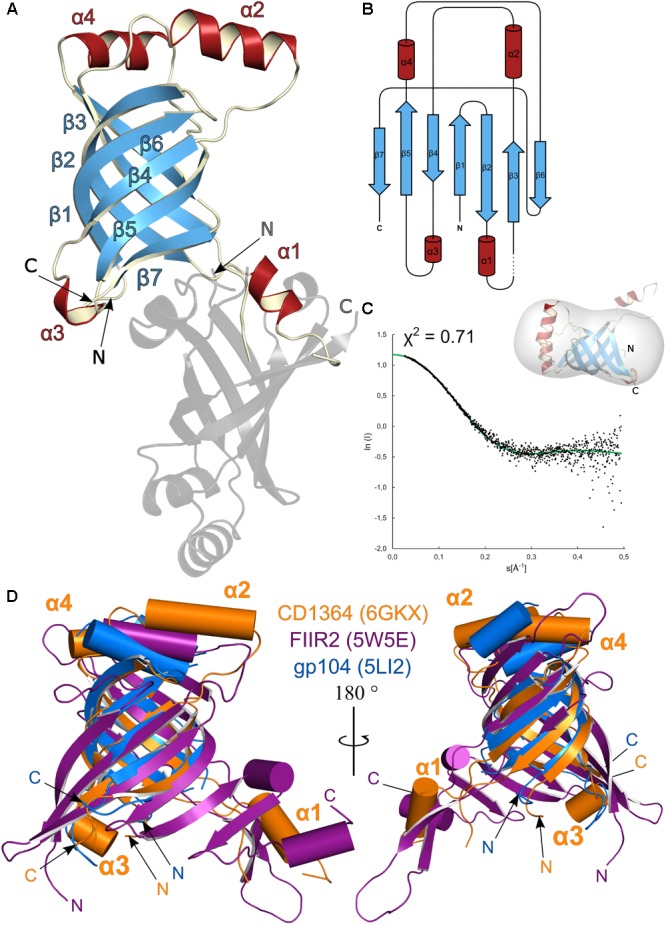
Structural analysis of Clostridium difficile R-type diffocin tube protein CD1364. The structures of monomeric CD1364 (blue and red) and a symmetry-related molecule (gray) are shown as cartoon representation in (A) and the corresponding topology diagram is shown in (B). Secondary structure motifs are numbered consecutively. SAXS analysis of purified CD1364 is presented in (C) with a fit of the experimental SAXS data (black dots) and a theoretical curve calculated from the crystal structure (green dots) together with the CD1364 crystal structure fitted to the calculated SAXS envelope. In (D), monomeric structures from homologous tube assembles of R-type pyocin (FIIR2; PDB: 5W5E; Zheng et al., 2017) and the bacteriophage φ812 tail (gp104; PDB: 5LI2; Nováček et al., 2016) were superimposed onto CD1364. N- and C-termini are labeled with N and C, respectively.