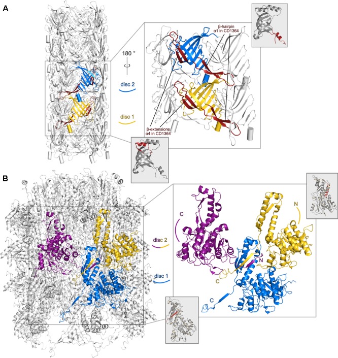
FIGURE 4.

Structural differences between monomeric R-type diffocin sheath and tube proteins CD1364 and CD1363 and the respective subunits in the R-type pyocin particle. Two subunits (FIIR2) from two disks of the tube (PDB: 5W5E; Zheng et al., 2017) are shown (blue and yellow) in cartoon presentation for pyocin in (A). A β-hairpin runs underneath a neighboring molecule of the same disk and establishes interactions with the molecules of one disk below. Each β-barrel is extended by two β-strands that form interactions with the adjacent central β-barrel. In (B), three subunits (FIR2) from two disks of the R-type pyocin sheath assembly (PDB: 3J9Q; Ge et al., 2015) forming a four-stranded β-sheet via exchanging and refolding of the N- and C-termini are shown (labeled with N and C, respectively). For comparison, structures of the monomeric unassembled R-type diffocin tube CD1364 and sheath CD1363 proteins are presented and regions that correspond to the respective R-type pyocin elements are indicated by red coloring.
