Figure 1.

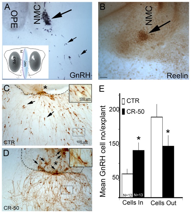
Reelin signaling blockade in vitro decreases gonadotropin releasing hormone-1 (GnRH) neuronal migration. (A) GnRH cells (blue-black, small arrows) have migrated into the periphery of the explant by 3 div, although large numbers remain at the tip of the nasal midline cartilage (NMC, large arrow). Inset: schematic of nasal explant (OPE, olfactory pit epithelium). (B) Immunostaining with antibody against Reelin in a 3 div explant, shows strong expression of Reelin (brown, arrow) at the tip of NMC. Reelin is expressed by mesenchymal cells in the area where GnRH neurons migrate into the periphery (Compare large arrows in A–D). 7 div explants immunostained with antibody against GnRH (brown) after chronic treatment (from 3–6 div) either with vehicle (CTR) (C) or CR-50 antibody (D), an antibody known to block the effect of Reelin. Dash lines indicate border between the main tissue mass and the periphery of the explant, asterisk indicates tip of NMC. Under control (CTR) conditions, GnRH-positive cells are dispersed outside the main tissue mass (C, arrows), in the periphery of the explants. After CR-50 treatment, the majority of the GnRH neurons remained within the main tissue mass (D, arrows) and fewer cells migrated into the periphery. (E) Quantitative analysis of GnRH cell distribution in the inner tissue mass and in the periphery under the two experimental conditions. The total number of GnRH cells did not change between control and treated explants. However, following CR-50 treatment significantly more GnRH neurons remained within the inner tissue mass (Cells In) as compared to control conditions (*P < 0.01; Student’s t-test). Concomitantly significantly less cells under CR-50 treatments were counted outside the main tissue mass (Cells Out) compared to CTR (*P < 0.01; Student’s t-test). Scale bar = 100 μm in (C) (same magnification A,D). Scale Bar = 50 μm in (D).
