. 2018 Jul 2;17:634–646. doi: 10.17179/excli2017-654

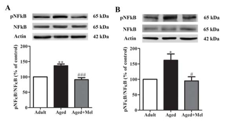
Figure 3. The effect of melatonin on pNFκB protein levels in the hippocampus (A) and the PFC (B) of aged mice. Mice were divided into three groups including the young control (2 months old), aged (22 months old) and melatonin-pretreated (10 mg/kg daily melatonin started from 16 to 22 months old) mice. Mice were sacrificed, brains were removed, hippocampus and prefrontal cortex were dissected and kept at -80 °C use. The protein levels of pNFκB and NFκB were determined by Western blot analyses. Representative bands from different groups are shown. The band densities were normalized to actin. The ratios were calculated as a percentage of the respective value of the control group. A one-way ANOVA was performed for statistical analysis. Data represent the mean ± S.E.M. from 4 mice. *p < 0.05 and **p < 0.01 compared with young adult mice, and #p < 0.05 and ###p < 0.001 compared with aged mice.

Figure 3