. 2018 Jul 4;17:647–662. doi: 10.17179/excli2018-1109

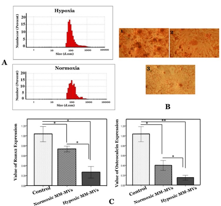
Figure 5. The effect of MM-MVS on osteoblasts differentiation. A) The scale bar shows size of MVs. B) Stained with alizarin red was conducted on 21 days. BM-MSCs were cultured in osteoinduction without MM-MVs (1), in the presence of 50 µg/ml MVs derived from hypoxic myeloma cells (2), and/or presence of 50 µg/ml MVs derived from normoxic myeloma cells (3). C) Results of Runx2 and osteocalcin expression in BM-MSCs that were cultured in osteogenic medium in the presence or absence of MM-MVs. RPMI-8226 cells were cultured in the normoxic and hypoxic chamber for the indicated periods (72 h). The MVs were harvested and added to osteogenic medium for 21 days. Quantification of these mRNA expressions normalized against the GAPDH mRNA levels. Data were obtained from three independent experiments and are presented as mean ± S.D. (* P <0.05, ** P <0.01).

Figure 5