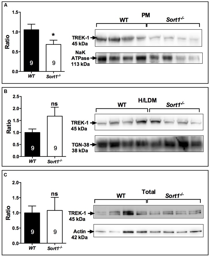
FIGURE 3.
Sub-cellular location of the TREK-1 channel protein in the brain of Sort1−/− and WT mice. (A) The TREK-1 expression was decreased by 36% (histogram, Left) in the plasma membrane (PM) prepared from Sort1−/− mouse brain when compared to PM prepared from WT mouse brain as visualized by Western blots (Right). (B) The expression of TREK-1 channels remained unchanged in high and low density vesicles (H/LDM) prepared from brains of Sort1−/− and WT mice, as well as in total brain extracts (C). The sub-cellular compartments were identified by specific markers: NaKATPase for plasma membranes (A), TGN38 for H/LDM (B), and actin for total brain extracts (C). ∗p < 0.05, ns, non-significant.

