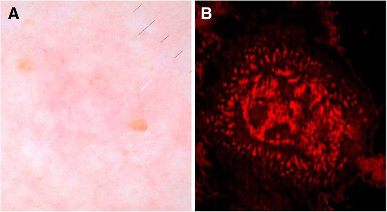
Fig. 4.

Dermoscopy and en face view of BCC on D-OCT: (a) Dermoscopy of BCC with pink-white shiny background, focal ulceration, arborizing vessels. b En face D-OCT of BCC shows disarray of thin, irregular vessels that are variable in size compared with the normal facial vessels. In comparison with more aggressive tumors, such as melanoma, the vascular pattern appears confined to the tumor. (Used with permission. Originally published by Levine, et al. 2017 in Optical Coherence Tomography in the Diagnosis of Skin Cancer)
