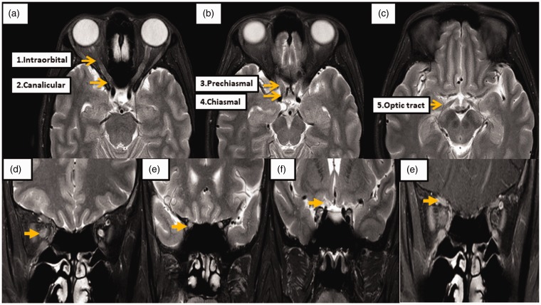
Figure 1.
Orbital magnetic resonance imaging (MRI) protocol and segmentations.
The orbital MRI findings were evaluated using 3-Tesla MRI (Philips, Ingenia). (a)–(c) Optic nerves are divided into the following five segments: intraorbital, canalicular, prechiasmal, chiasmal, and optic tract. The T2-weighted image with fat-suppression technique was used for evaluating signal intensity and diameter of the optic nerve. The optic nerves were measured at (d) mid-part of the intraorbital segment, (e) canalicular segment and (f) prechiasmatic segment, and (g) optic nerve enhancement is demonstrated on a T1-weighted image postcontrast study with fat-suppression techniques at the intraorbital part of the right optic nerve.

