Figure 1.

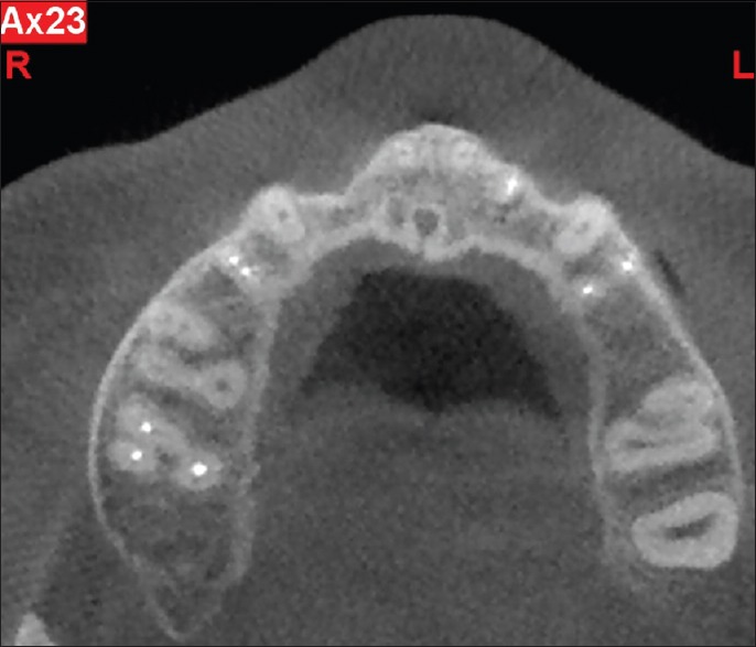
Axial cone beam computed tomography view shows two distinct canals in the mesiobuccal root of the maxillary right first molar tooth

Axial cone beam computed tomography view shows two distinct canals in the mesiobuccal root of the maxillary right first molar tooth