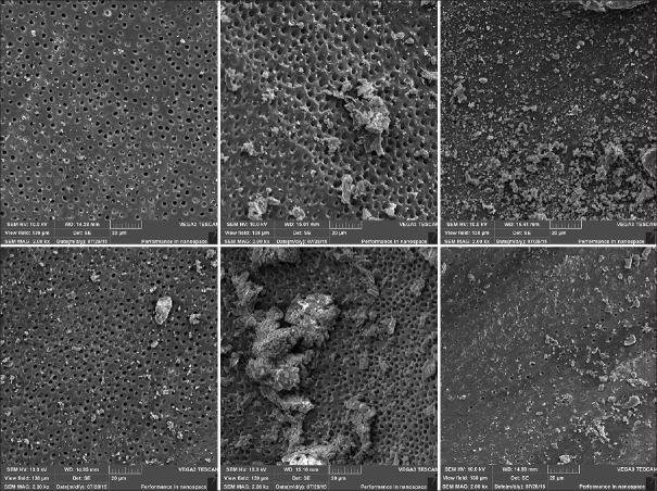
Figure 1.
Scanning electron microscope images representative of the root canal walls in the mesiodistal (above) and buccolingual (below) directions after instrumentation with the HyFlex system. Please observe the similar quantity of smear layer adhered in both directions of analysis, and in the root canal thirds (cervical-left; middle-center; and apical-right) (×1000)

