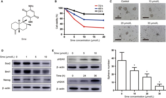
Figure 2.

Sme preferentially inhibits MCF7‐Nanog cells proliferation and caused DNA damage. A, The chemical structure of Sme. B, Cytotoxicity of Sme to MCF7‐Nanog cells. Cells were incubated with Sme with different concentrations for indicated time points, and the viability was determined by CCK‐8 assay. C, MCF7‐Nanog cells were exposed to Sme for 72 hours and subjected to sphere‐forming assay. Scale bar, 100 μm. Data are presented as mean ± SD. * P < .05. D, Sme suppressed the expression of stemness‐related markers in MCF7‐Nanog cells. Western blotting was used to detect the expression of Nanog, Sox2, and Bmi1 after treatment with Sme for 36 h. E, The expression of γH2AX was decreased in dose‐ and time‐dependent manner after exposure to Sme
