Figure 6.

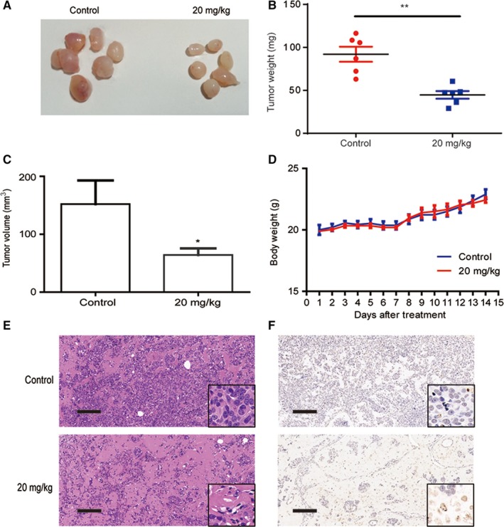
Sme inhibited tumor growth in vivo. MCF7‐Nanog cells were injected into the mammary fat pad of female NOD/SCID mice. Two weeks after injection, the mice were randomly divided into two groups: control and Sme (20 mg/kg), followed by intraperitoneally injection with Sme every other day for two weeks. A, The tumors were surgically excised and photographed two weeks after Sme treatment. B, C, The tumor weight and volume were expressed as mean ± SD. * P < .05, ** P < .01. D, The body weight was monitored every day after Sme treatment and expressed as mean ± SD. E, The histology was assessed by H&E staining. Scale bar, 100 μm. F, TUNEL assay was used to evaluate apoptotic status of tumor tissues. Scale bar, 100 μm
