
Figure 1. Detergent treatment after plasmolysis causes further contraction of the Gram-negative cell wall.
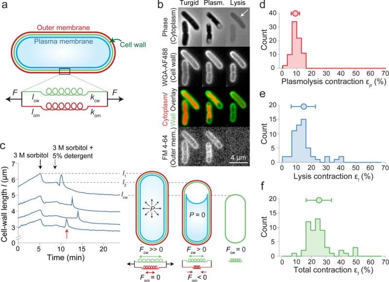
a) Model of the cell wall/outer membrane complex as parallel linear springs with spring constants kcw, kom, and rest lengths lcw, lom.
b) E. coli cells (turgid, plasmolyzed, lysed) stained with WGA-488 and FM 4-64. White arrow: residual phase signal after lysis (n = 84 cells, 3 experiments).
c) Left: cell-wall length versus time during hyperosmotic shock and treatment with detergent for representative cells (n = 79 cells). Red arrow: sharp swelling upon lysis. Right: model of turgid/plasmolyzed/lysed cellular state.
d-f) Histograms of length contraction upon (d) plasmolysis (n = 79 cells), (e) lysis (n = 56 cells), and (f) in total (n = 56 cells). Circle and error bars, mean ± 1 s.d.