Figure 2.

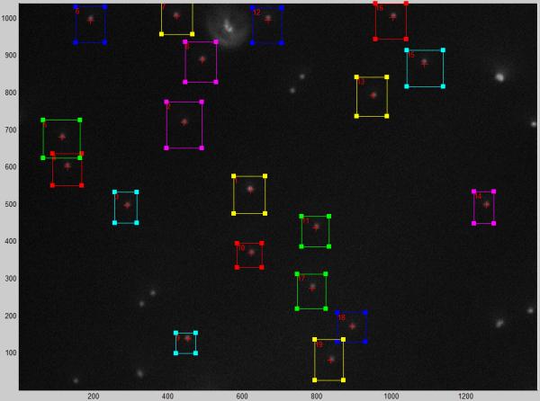
A screen shot from the microscope control program with regions of interest (boxes) superimposed on a dark field image of 320 nm, tethered particles. The cursors update continuously within the stationary boxes in the live image.

A screen shot from the microscope control program with regions of interest (boxes) superimposed on a dark field image of 320 nm, tethered particles. The cursors update continuously within the stationary boxes in the live image.