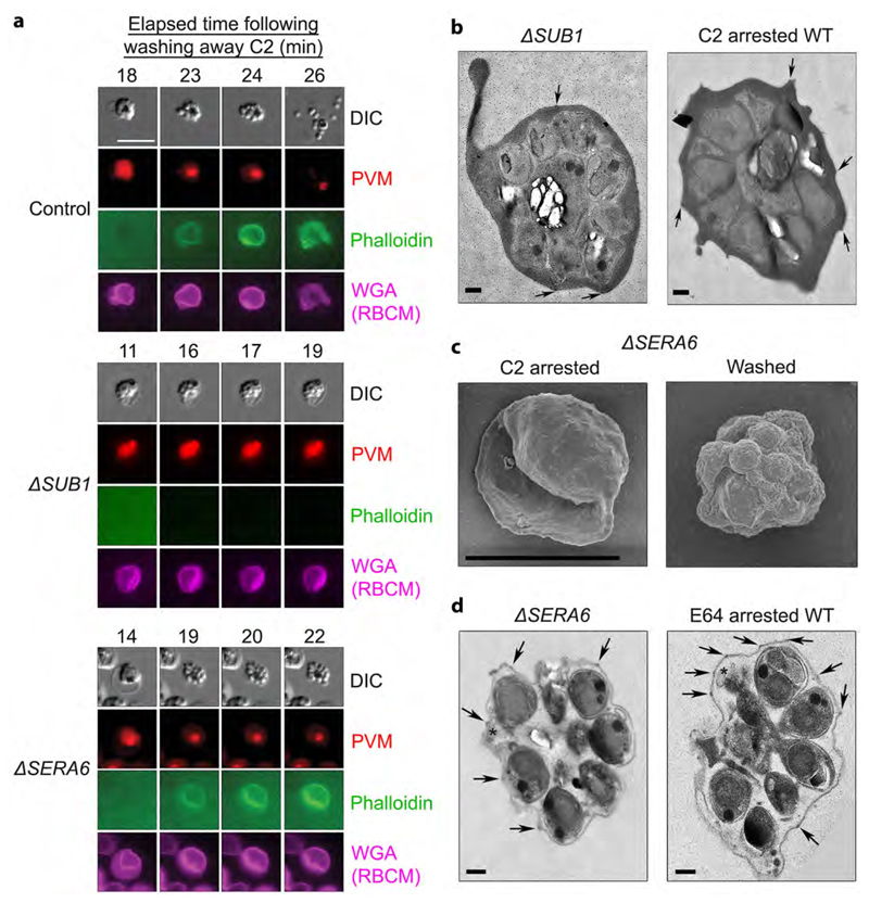
Figure 3. SUB1 is required for PVM disruption and RBCM poration, whereas the ΔSERA6 phenotype mimics egress arrest with the cysteine protease inhibitor E64.
a, Stills from simultaneous time-lapse DIC and fluorescence microscopic examination of typical control WT, ΔSUB1 and ΔSERA6 schizonts at the indicated intervals following removal of the egress inhibitor C2. PVM rupture and RBCM poration (indicated by access of phalloidin to the host RBC cytoskeleton) occurs in the ΔSERA6 parasites but not in the ΔSUB1 parasites, whilst RBCM rupture occurs in neither mutant. Scale bar, 10 μm. b, TEM micrographs of an arrested ΔSUB1 schizont and a C2-arrested control cell, showing that the trapped merozoites are surrounded in both cases by an intact PVM and RBCM. Knob structures characteristic of the parasite-infected RBCM6 are indicated on its outer surface (arrow heads). The black dots are gold fiducials added for tomography. Scale bar, 500 nm. c, SEM images of ΔSERA6 schizonts before and 30 min following C2 removal, showing collapse of the RBCM around the intracellular merozoites in the washed sample. Scale bar, 5 μm. d, TEM micrographs of an arrested ΔSERA6 schizont and an E64-arrested control cell, showing in both remnants of ruptured PVM (asterisks) adjacent to the trapped merozoites. Knobs are highlighted as above (arrow heads). Scale bar, 500 nm. All experiments were repeated twice, with reproducible results.

