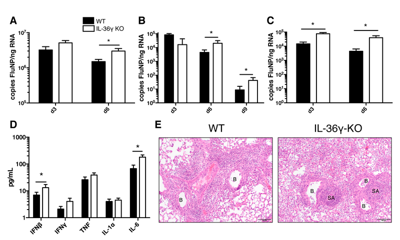
Figure 3: Viral titers, cytokine levels, and pathology of WT and IL-36γ-KO mice following influenza infection.
Age- and sex-matched C57Bl/6J (filled bars) and Il36g−/− mice (open bars) were infected with (A) influenza PR8, (B) 30,000 EID50 ×31, or (C) 300,000 EID50 ×31 and lungs were analyzed for influenza NP copy number. (D) Bronchoalveolar lavage fluid was analyzed for expression of proinflammatory cytokines on day 3 post-infection in C57Bl/6J (filled bars) and Il36g−/− mice (open bars). (E) Example images of formalin-fixed, paraffin-embedded, H&E-stained lungs from C57Bl/6J and Il36g−/− mice. Inflamed bronchioles (B) and small arterioles (SA) are noted on the images. For A-D, data are combined from 2–3 independent replicate experiments of at least 5 mice per group per replicate. Males and females were used in equal proportions. Asterisks indicate significant differences by Student’s t test on log-transformed data.

