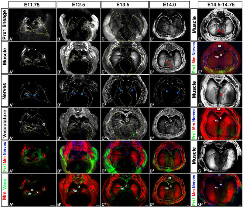
Figure 6.
Morphogenesis of PPF-derived muscle connective tissue and central tendon, muscle, phrenic nerve, and vasculature are tightly linked temporally and spatially. (A) Muscle progenitors (A2) and axons of the phrenic nerve (A3) migrate into the right PPF (A1) adjacent to the vena cava (vc) and into the left PPF and caudal to the vestigial left horn of the sinus venosus (*). Vasculature (A4) also forms in PPF region in close association with vena cava and left sinus venosus horn. (B–D) PPFs migrate dorsally and ventrally to cover the surface of the underlying liver. In close association with the PPFs, muscle and vasculature expands as well as phrenic nerve axons extend dorsally and ventrally. Vessels from the body wall merge with the diaphragm’s vasculature (green arrow, C4). (D–G) Muscle progenitors appear and differentiate into myofibers in the central tendon region (D–F), but are gradually removed (G). nt, neural tube; e, esophagus; vc, vena cava. Scale bars A – G = 200um. 3-dimensional movies in Supplemental Movies 13–16.

