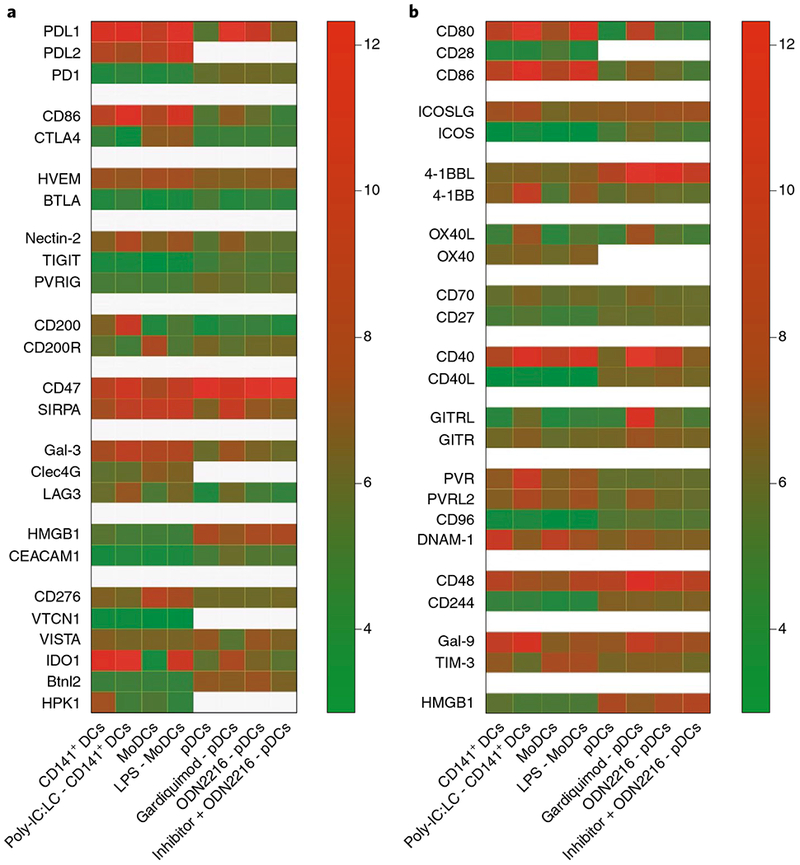
Fig. 2 |. Expression of selected immune checkpoint factors on in vitro stimulated CD141+ DCs, MoDCs and pDCs.

Different DC subsets display a unique pattern of expression for inhibitory and activating checkpoints both at basal levels and on activation. CD141+ DCs and MoDCs were differentiated from CD34+ precursors and monocytes, respectively41. Natural pDCs were sorted from blood of healthy donors42. The original microarray datasets were analysed using the statistical package limma in R. Expression values are normalized by housekeeping gene expression43, and were log2 transformed; mean values are shown on the heatmap. a, Inhibitory immune-checkpoint interactions. b, Stimulatory immune-checkpoint interactions. LPS, lipopolysaccharide-TLR4 agonist; Gardiquimod, TLR7 agonist; ODN2216, Class A CpG oligonucleotide-human TLR9 ligand.
