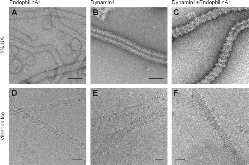
Figure 5: Membrane-remodeling complexes visualized with negative stain versus cryo-electron microscopy.
EndophilinA1 (A and D), Dynamin1 (B and E), or the hetero-complex formed by both proteins (C and F). In all cases, the purified proteins remodel spherical vesicles into high-curvature cylinders wrapped in an oligomeric protein coat. In the hetero-complex the adjacent turns of the Dynamin1 helix are separated by 2, 3 or 4 copies of an interleaving molecule of EndophilinA1. Bars 50nm.

