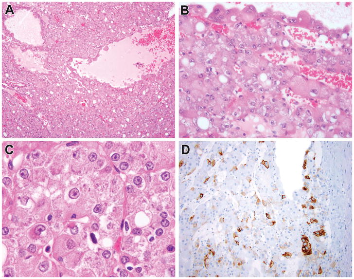
Figure 2.
Previously reported ESC RCC that lacked detectable TSC mutations. This tumor corresponds to ESC RCC #10 of reference 5, multifocal cystic neoplasms in a 14-year-old female with a history of sickle cell trait. This neoplasm demonstrated solid and cystic areas and cells with voluminous cytoplasm lining the cysts, typical of ESC RCC (A, B). On high power examination, the neoplastic cells had granular pink cytoplasm with granular cytoplasmic stippling, characteristic of ESC RCC (C). The neoplasm demonstrated patchy immunoreactivity for cytokeratin 20 (D), again typical of ESC RCC. While this neoplasm demonstrated all of the hallmark features of ESC RCC, it lacked demonstrable mutations in TSC1 and TSC2.

