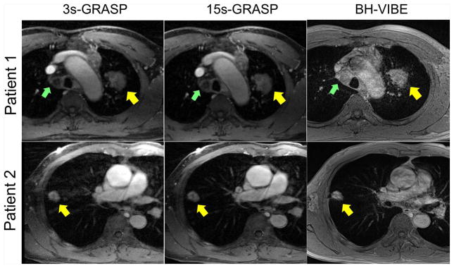
Figure 5.
Post-contrast phases in two patients comparing GRASP with BH-VIBE images. One patient (top row) was diagnosed as squamous cell carcinoma and the other patient (bottom row) was diagnosed as adenocarcinoma. Although the 3s-GRASP images show certain residual artifacts with relatively lower image quality due to high acceleration rate, the 15s-GRASP images yielded overall image quality that is comparable with BH-VIBE and with less motion artifacts. In particular, the mediastinal lymph node in patient 1 (indicated by green arrows) was not clearly delineated in the BH-VIBE images due to motion-induced artifacts in great vessels.

