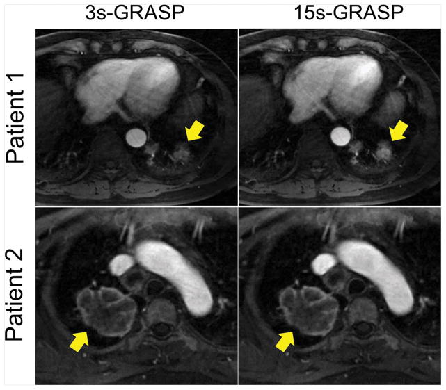
Figure 6.
A comparison of the arterial phase between 15s-GRASP and 3s-GRASP in two patients. In patient 1, the lesion (yellow arrows) is relatively smaller and closer to the diaphragm, and thus the 15s-GRASP image shows better lesion delineation than the 3s-GRASP image. However, they both show good delineation of the mass in patient 2.

