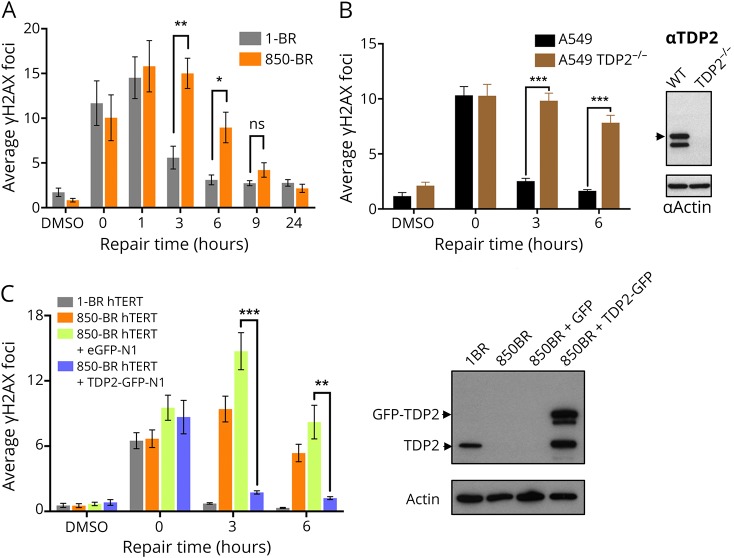
Figure 3. Reduced DSB repair in TDP2-mutated patient fibroblasts and A549 cells after topoisomerase 2-induced DNA damage.
(A) DSBs were measured by γH2AX immunostaining in normal 1-BR and patient 850-BR primary fibroblasts before and after treatment for 30 minutes with DMSO vehicle or 25 μM etoposide (“0”), followed by subsequent incubation in a drug-free medium for the indicated repair periods. (B) DSBs were measured as above, in wild-type A549 cells and in A549 cells in which TDP2 was deleted by CRISPR/Cas9 gene editing (TDP2−/− A549). The level of TDP2 and actin (loading control) in the cell lines used for these experiments is shown by Western blotting on the right. (C) DSBs were measured as above in hTERT-immortalized 1-BR fibroblasts and 850-BR fibroblasts and in the latter cells after transfection with the empty GFP vector or vector encoding recombinant human TDP2-GFP. The level of TDP2 and actin (loading control) in the cell lines used for these experiments is shown by Western blotting on the right. Data are mean ± SEM of 3 independent experiments, and statistically significant differences were determined by the t test (ns = not significant, *p < 0.05; **p < 0.01; ***p < 0.001). DSB = double-strand break; GFP = green fluorescent protein; TDP2 = tyrosyl DNA phosphodiesterase 2.

