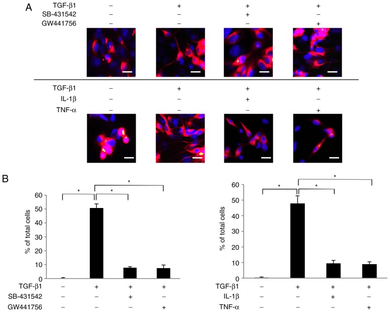
Figure 4.
Nerve growth factor secreted by SCDC2 cells following TGF-β1 stimulation promoted neurite extension from the surface of ATPγS-treated PC12 cells. (A) Neurite extension of PC12 cells was visualized by immunostaining (×200 magnification; scale bar, 50 µm) with anti-neurofilament H antibody (red) and nuclei were stained with DAPI (blue). SCDC2 cells (2×10⁴ cells) and rat pheochromocytoma cells PC12 (1×10⁴ cells) were co-cultured and treated with or without TGF-β1 (10 ng/ml) for 4 days. Cells were also treated with TGF-β type I receptor inhibitor SB-431542 (10 µM), TrkA inhibitor GW441756 (2 nM), IL-1β (10 ng/ml), or TNF-α (10 ng/ml) from the beginning of the co-culture. In addition, ATPγS (100 µM) was added to all cultures during cell seeding. Dimethyl sulfoxide was added to cell cultures as a vehicle control for SB-431542 and GW441756, respectively. (B) Statistical assessment of neurite extension in PC12 cells co-cultured with SCDC2 cells. Data represent the mean ± standard deviation (n=8). *P<0.05. TGF, transforming growth factor; SCDC, single cell-derived culture; IL, interleukin; TNF, tumor necrosis factor; ATPγS, adenosine 5′-O-(3-thio)triphosphate; TrkA, tropomyosin receptor kinase A.

