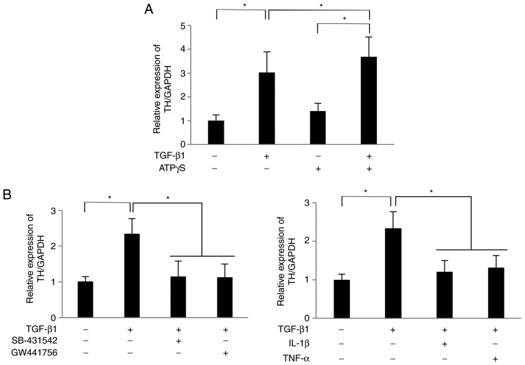
Figure 5.
Nerve growth factor secreted by SCDC2 cells subsequent to TGF-β1 stimulation promoted the expression of TH mRNA in PC12 cells. SCDC2 cells (7×10⁴ cells) and rat pheochromocytoma cells PC12 cells (3.5×10⁴ cells) were co-cultured and stimulated with or without TGF-β1 (10 ng/ml) for 24 h. The relative expression level of TH was evaluated using reverse transcription-quantitative polymerase chain reaction in cells also treated with (A) ATPγS (100 µM), and with (B) TGF-β type I receptor inhibitor SB-431542 (10 µM), TrkA inhibitor GW441756 (2 nM), IL-1β (10 ng/ml) or TNF-α (10 ng/ml) during the co-culture. Dimethyl sulfoxide was added to cell cultures as a vehicle control for SB-431542 and GW441756, respectively. Data represent the mean ± standard deviation (n=6). *P<0.05. TGF, transforming growth factor; SCDC, single cell-derived culture; IL, interleukin; TNF, tumor necrosis factor; ATPγS, adenosine 5′-O-(3-thio)triphosphate; TrkA, tropomyosin receptor kinase A; TH, tyrosine hydroxylase.

