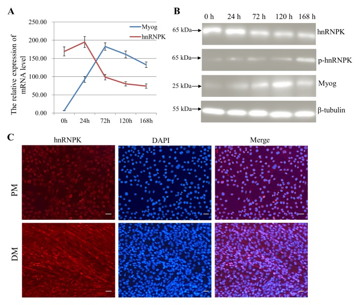
Fig. 1.
hnRNPK is upregulated during C2C12 myogenesis. (A) qRT-PCR analysis was performed on hnRNPK and Myog from C2C12 cells at different times post differentiation. Gapdh was used as an internal control. (B) Western blot analysis of hnRNPK was performed in C2C12 cells grown in growth medium, as well as after 24, 72, 120 and 168 h in DM. (C) hnRNPK localisation changes in C2C12 myoblasts during differentiation. Cells were proliferating (PM) or differentiating for 72 h (DM). Cellular hnRNPK (red) was detected by indirect IF. Cells were DAPI-stained to reveal DNA (blue) prior to confocal fluorescence microscopy. Scale bar = 100 μm.

