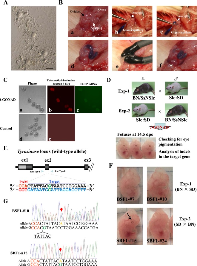
Figure 1.
(A) One-cell rat embryos isolated from 0.7 dpc female SD rats. Note the presence of cumulus cell-free 1-cell embryos. Bar = 100 μm. (B) The i-GONAD procedure. Under appropriate anesthesia, ovary/oviduct/uterus of a pregnant rat at 0.7 dpc was exposed under a dissecting microscope. After positioning the oviduct, it was gently grasped in forceps (a). A micropipette was then inserted into the lumen of the oviduct by piercing the oviduct wall and (controlled by a mouthpiece) approximately ~1.5 µL solution was immediately expelled into the oviduct (b). The injected substances can be easily identified by the co-injected Fast Green (c,d). After instillation, the entire oviduct was covered with a piece of wet Kimwipe towel and then subjected to in vivo electroporation using tweezer-type electrodes (e). After removal of electrodes, the electroporated area remains intact (f). (C) Detection of fluorescent 2-cell embryos after i-GONAD. When pregnant (0.7 dpc) SD rats were subjected to in vivo electroporation after intraoviductal instillation of Rhodamine + EGFP mRNA-containing solution (i-GONAD), some of the recovered 2-cell embryos exhibited distinct red and/or green fluorescence (a–c). In contrast, intraoviductal instillation of Rhodamine alone (without electroporation) (Control) resulted in embryos showing no fluorescence (d,e). Bar = 100 μm. (D) Flowchart of experiments to test the feasibility of i-GONAD for the production of mid-gestational fetuses carrying indels at a single target locus (Tyr). In Exp-1, BN females were mated to SD males to obtain pregnant BN rats, while in Exp-2, SD females were mated to BN males to obtain pregnant SD rats. (E) Schematic illustration of the wild-type Tyr locus. The target sequence of Tyr exon 2 (ex2) recognized by gRNA is overlined and the PAM sequence is marked in red. The target nucleotide “G” marked in green is a key nucleotide for tyrosinase activity; nucleotide replacement at this position often causes an albino phenotype. Primers for amplification 598 bp of exon 2, Rat Tyr-F and -R, are shown. (F) Offspring obtained after i-GONAD in Exp-1 or -2. The embryos numbered BSF1-#7, BSF1-#10 and SBF1-#24 exhibited non-pigmented eyes. The fetus numbered SBF1-#15 had pigmented eyes (shown by an arrow), probably as a result of unsuccessful genome editing at the Tyr locus. (G) Direct sequencing of PCR products derived from the embryos shown in (F). Red arrow in BSF1-#10, which showed eye non-pigmentation, indicate heterozygous bi-allelic KO at the Tyr target (nucleotide “G” marked in green in E) because one allele (called allele-A) has the nucleotide “A” (shown in brown) and the other allele (called wild-type allele or allele-G) lacks 6-bp sequence above the PAM. In contrast, the red arrow in SBF1-#15 indicates heterozygous mono-allelic KO, because one allele (called allele-A) has the nucleotide “A” (shown in brown), whereas the wild-type nucleotide “G” exists at the same position as allele-G.

