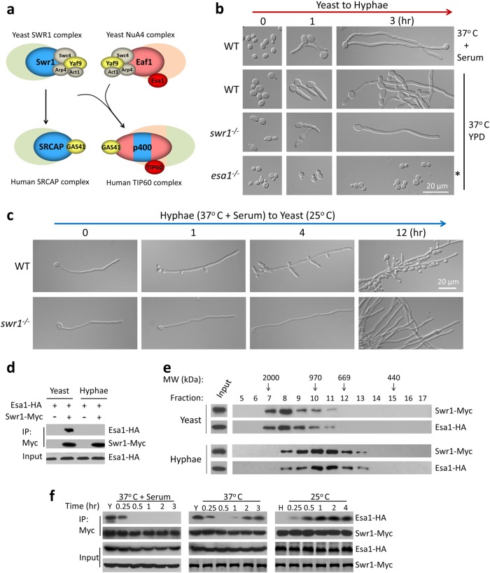
Fig. 1. Merge and separation of NuA4 and SWR1 complexes are correlated with the reversible yeast–hypha transition in Candida albicans.
a Schematic diagram showing a proposed model for the evolution of NuA4 and SWR1 complexes from yeast to human. b Swr1 and Esa1 have opposite roles during hyphal development. An overnight culture of wild-type (SC5314) yeast cells were diluted 1:100 into fresh YPD medium and induced at 37 °C in the presence or absence of 10% serum. The swr1 mutant was induced at 37 °C and esa1 mutant at 35 °C (asterisked) in YPD without serum. Cell morphoplogy was observed at 0 h, 1 h and 3 h. d Hyphae-to-yeast transition. The induced hyphae of wild-type and swr1 mutant (in YPD with 10% serum at 37 °C for 3 h) were re-cultured in fresh YPD at 25 °C for indicated time. d, e Esa1 and Swr1 associated in yeast and dissociated in hyphae. A wild-type strain (BWP17) carrying Esa1-HA and Swr1-Myc under their endogenous promoters was cultured in YPD at 25 °C for 12 h to OD600 ~3 (yeast state) or YPD with 10% serum at 37 °C for 3 h (hyphae state). Whole-cell extracts (WCEs) of yeast or hyphae were subjected for co-immunoprecipation (Co-IP) (d) or Gel filtration (e) experiments. WCEs were immunoprecipitated with anti-Myc antibody and probed with anti-HA or anti-Myc (d). Every second fraction eluted from a Superose 6 column was analyzed for the presence of Swr1-Myc, together with Esa1-HA by western blotting (e). Native molecular weight markers eluting in the corresponding fractions are indicated on the top of the panel. f Esa1-Swr1 association during reversible yeast–hypha transition. The cells were cultured in conditions described and collected at time points indicated for Co-IP. Western blot analysis was carried out using a peroxidase-conjugated anti-Myc antibody or anti-HA antibody to assess levels of Swr1-Myc or Esa1-HA

