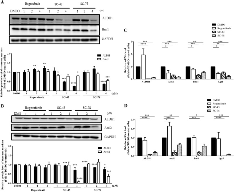
Fig. 4. Effects of regorafenib, SC-43, and SC-78 on the expression of stemness markers in both HCT-116 and HT-29 cells.
Total lysates (20 μg) were prepared from (a) HCT-116 and b HT-29 cells after they were treated with DMSO (vehicle) or different doses (1, 2, and 4 µM) of regorafenib, SC-43, and SC-78 for 72 h and subjected to western blot analyses using primary antibodies against ALDH1 and Bmi1 (for HCT-116 cells) or ALDH1 and Ascl2 (for HT-29 cells). GAPDH was used as a loading control. The quantitative results obtained by densitometry are the mean ± SD of three independent experiments (lower panels). *p < 0.05, **p < 0.01, ***p < 0.005, and ****p < 0.001 compared with DMSO-treated cells by Student’s t-test. Total RNA samples (5 μg) were isolated from c HCT-116 and d HT-29 cells after they were treated with DMSO or 4 µM of regorafenib, SC-43, and SC-78 for 48 h and subjected to qRT-PCR analyses to determine the mRNA levels of several CRCSC marker genes. Data are the mean ± SD of three independent experiments. *p < 0.05, **p < 0.01, ***p < 0.005, and ****p < 0.001 compared with DMSO-treated cells by Student’s t-test

