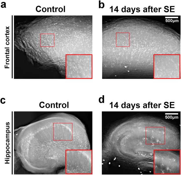
Figure 3.
Representative X-ray absorption projections of Golgi-Cox labeled brain structures. X-ray projection of a hemi-frontal cortex from control (a) or pilocarpine-treated animals. (b) X-ray projection of the medium part of a hippocampus from control (c) or pilocarpine-treated animals (d). Regions of interest show in detail the mercury-impregnated neurons with typical cell body and neurites. Inserts from (c) and (d) show a significant reduction on total number of hippocampal neurons together with an alteration on cell branching and distribution.

