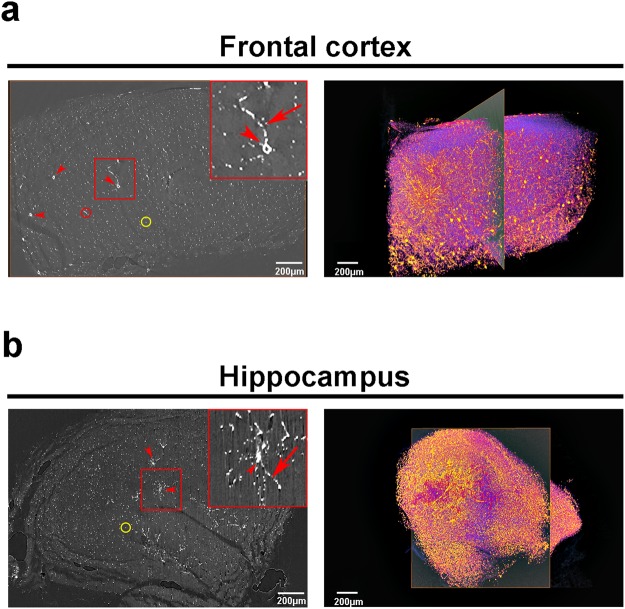
Figure 4.
Synchrotron X-ray microtomography of mercury-impregnated neurons allows virtual slicing and 3D rendering of whole brain structures. Reconstructed slice (left panel) and 3D image rendering (right panel) of a control frontal cortex (a) and hippocampus (b). Rectangles in (a) and (b) highlights, respectively, a single cortical or hippocampal neuron. Arrowheads: neuron cell bodies; arrows: neurites; red circles: longitudinal virtual slicing of a neurite; yellow circles: transversal virtual slicing of a neurite.

