Fig. 5.
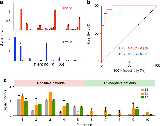
Molecular profiling of patient samples. a HPV 16 and HPV 18 signals were measured from clinical endocervical brush samples (n = 35). The L1-specific signals are shown for comparison with the clinical gold standard. See Supplementary Fig. 14 for multi-loci measurements on all clinical specimens. b Receiver operator characteristic (ROC) curves of the HPV 16 and HPV 18 L1 locus assays were used to determine the detection accuracies. HPV 16 assay showed 92.9% sensitivity (13/14) and 90.5% specificity (19/21) and HPV 18 assay showed 83.3% sensitivity (5/6) and 100% specificity (29/29) at the Youden’s index cutoff. c Locus-specific HPV 16 enVision assays (L1, L2, and E1 locus assays) were performed in all patients. Representative examples from L1-positive (left) and L1-negative (right) patients are shown. Note that in the subset of L1-negative patients, the inclusion of L2 and E1 locus assays could improve the detection coverage to identify previously undetectable infections. This was further validated through independent Taqman fluorescence analysis, which showed a high concordance with the enVision results in all tested clinical specimens (see Supplementary Fig. 15). All measurements were performed in triplicate, and the data are displayed as mean ± s.d. AUC, area under the curve

