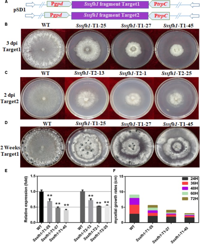
FIGURE 2.

Biological characterization of the Sssfh1-silenced transformants. (A) Diagrams of constructs used to silence Sssfh1 in S. sclerotiorum. (B,C) The hyphal growth of WT, Sssfh1-Target1-silenced strains and Sssfh1-Target2-silenced strains. Selected Target1 transformants and WT strain were inoculated on PDA plates and the pictures were taken 3 days post-inoculation (dpi) at 25°C, and the pictures of Target2 transformants were taken 2 dpi. (D) Sclerotial morphology of the WT and RNAi strains. Transformants and WT strain were cultured on PDA plates and grown at 25°C. Pictures were taken 14 dpi. (E) Relative expression level of Sssfh1 in different RNAi strains. Transformants and WT strain were inoculated on PDA media for two days and the hyphae were collected for RNA preparation. A value of one was assigned to the relative abundance of Sssfh1 in the WT strain. The gene expression levels of Sssfh1 in the silenced transformants and the WT strain were normalized to that of the mean of tubulin, actin, and histone transcripts from each strain. Data = means ± SD, one-way ANOVA, ∗∗indicates significance at p < 0.01. (F) Hyphal growth rates of the RNAi strains and the WT strain cultured on PDA plates. The first measurement began at 24 hpi, and the interval was set as every 12 h. The measurement was completed at 72 hpi. Three independent biological replications were performed.
