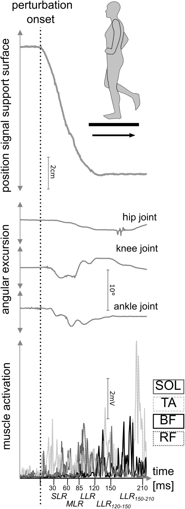
FIGURE 2.
Outcome measures during posterior perturbation of a representative participant. Recording of neuromuscular data (bottom) and kinematic data (middle) during posterior translation of the support surface (top) were carried out. For muscular activation, the soleus (SOL), tibialis anterior (TA), biceps femoris (BF), and rectus femoris (RF) muscles were measured during short- (SLR), medium- (MLR), and long-latency responses (LLR) after perturbation onset. Simultaneously, joint excursions of the ankle, knee, and hip joint were assessed.

