
FIGURE 6.
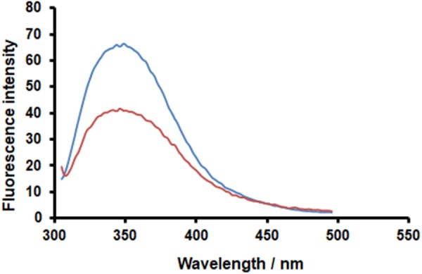
PAC-A2 determines quenching of HA-IAV fluorescence. Recombinant HA-IAV was resuspended in PBS buffer (pH 7.4) at a concentration of 1.2 μM. Fluorescence of HA-IAV aliquots were measured in the absence (blue curve) and presence (orange curve) of 25 M PAC-A2 after incubation for 1 h at 25°C. Fluorescence emission spectra were monitored in the 295–500 nm range using a scan-rate of 200 nm/min upon excitation at 290 nm. Data reported were normalized by subtracting the PAC-A2 fluorescence contribution.