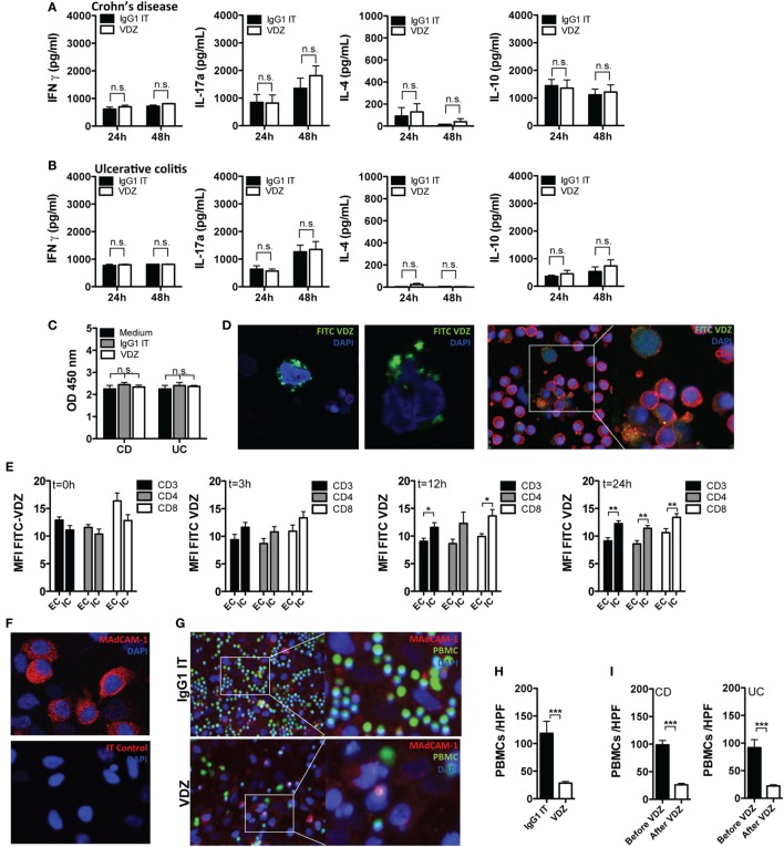
Figure 4.
Cellular mechanism of action of vedolizumab (VDZ). (A,B) Peripheral blood leukocytes from patients with Crohn’s disease (CD) [(A), n = 7] and ulcerative colitis (UC) [(B), n = 7] were incubated with VDZ or isotype (IT) control for 24 and 48 h. Afterward, concentration of IFNγ, IL-4, IL17a, and IL-10 in the supernatant was quantified by ELISA. (C) Peripheral blood leukocytes from patients with CD (n = 7) and UC (n = 7) were incubated with VDZ, IT, or media only for 24 h. Afterward, release of cytoplasmic histone-associated DNA fragments as a marker of induced cell death was quantified by ELISA. (D) Confocal microscopy of peripheral blood leukocytes (left panels) and sorted CD4+ T cells (right panels) incubated for 24 h with fluorescein isothiocyanate (FITC)-labeled VDZ. (E) Internalization of α4β7 after binding to VDZ over time. Peripheral leukocytes from seven inflammatory bowel disease patients were incubated for the indicated time points with VDZ. From each patient, 5 × 10⁵ peripheral leukocytes were first permeabilized to make the intracellular compartment accessible for staining followed by staining with fluorescent labeled VDZ. Another 5 × 10⁵ cells were first stained with FITC-VDZ and were then permeabilized. Afterward, α4β7 expression was quantified in CD3+, CD4+, and CD8+ T cells. (F) Mucosal addressin cell adhesion molecule-1 (MAdCAM-1) expression in HeLa cells. Upper panel: staining of MAdCAM-1. Lower panel: isotype control. (G,H) Peripheral blood leukocytes from healthy controls (n = 4) were labeled with a vital dye (green) and co-incubated with IgG1 and VDZ for 60 min on confluent grown MAdCAM-1 expressing HeLa cells (red). Afterward, non-attaching cells were removed by repeated washing. Adhering cells were visualized with fluorescence microscopy and attaching leukocytes per high power field (HPF) were counted. (I) Peripheral leukocytes from CD (n = 3) and UC (n = 3) patients before and after completion of VDZ induction therapy from the same patient were labeled with a vital dye and co-incubated with IgG1 and VDZ for 60 min on confluent grown MAdCAM-1 expressing HeLa cells. Afterward, non-attaching cells were removed by repeated washing. Adhering cells were visualized with fluorescence microscopy and attaching leukocytes per HPF were counted. Results are presented as mean ± SEM. Differences between samples are compared with the Mann–Whitney U test (*p < 0.05; **p < 0.01; ***p < 0.001).

