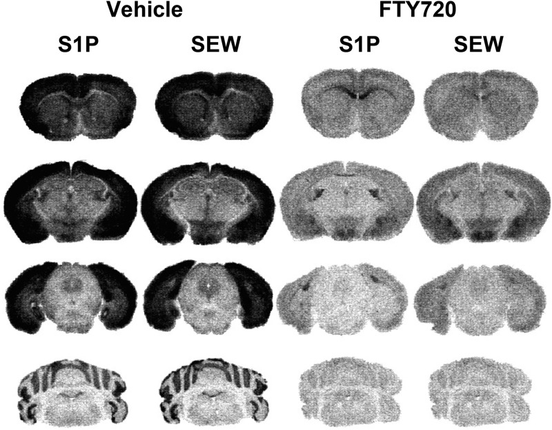
Fig. 8.
Agonist-stimulated [35S]GTPγS autoradiography reveals widespread loss of S1PR-mediated G protein activation in the brain in repeated FTY720-treated mice. Mice were treated with FTY720 (2 mg/kg, twice per day) or vehicle for 6.5 days, and brains were processed for S1P (60 µM)- or SEW2817 (80 µM)-stimulated [35S]GTPγS autoradiography. Representative images from mice treated with vehicle or FTY720 (n = 7 to 8 mice per group) are shown in caudate-putamen, cingulate cortex, and corpus callosum (row 1); hippocampus, amygdala, hypothalamus, and stria terminalis (row 2); PAG (row 3); and cerebellum (row 4).

