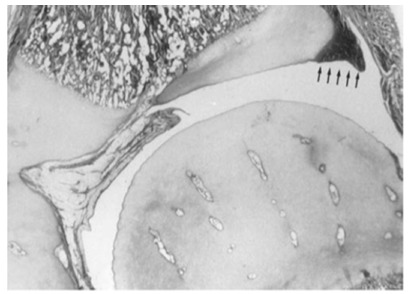
Fig. 3.

Hip joint in an infant. Note the vascular channels in the cartilaginous femoral head and the acetabular cartilage and labrum at the periphery (reproduced with permission from Weinstein SL, Flynn JJ, eds. Lovell and Winter’s Pediatric Orthopaedics. Vol. 2. Seventh ed. Philadelphia: Lippincott Williams & Wilkins, 2014).14
