
Fig. 2.
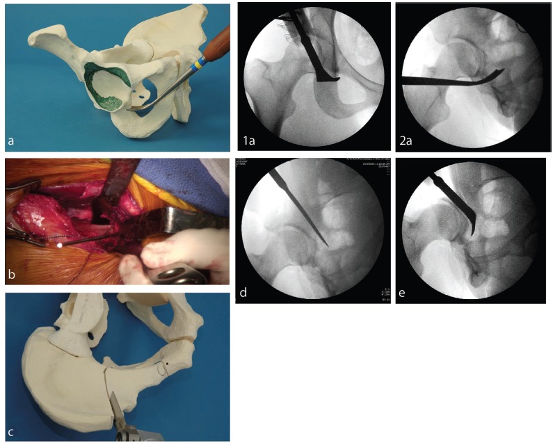
Intraoperative images confirm desired placement of osteotomies and desired realignment of the acetabulum to compensate for both anterior and lateral insufficiency: (a) anterior ischial osteotomy (infracotyloid groove), showing (1) correct placement within the groove, just distal to inferior lip of acetabulum and (2) image intensifier in anteroposterior and oblique projections confirms correct placement of chisel just distal to acetabulum; (b) superior pubic ramus osteotomy lies just medial to iliopectineal eminence; (c) iliac osteotomy begins just distal to the anterior superior spine, directed toward the apex of the greater sciatic notch. It ends at a point about 1 cm lateral to the iliopectineal line and approximately 3 cm anterior to the sacroiliac joint; (d) the posterior column osteotomy is made with a straight chisel. It begins at the posterior end of the iliac osteotomy. It is directed at the ischial spine, bisecting the posterior column, safely between the posterior acetabular wall and the anterior wall of the greater sciatic notch. The image intensifier in an oblique projection can confirm the proper position of this osoteotomy; (e) posterior ischial osteotomy. This osteotomy is made with a curved or angled chisel.