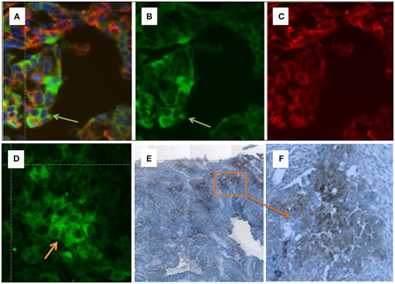
Figure 4.
(A–F) Immunohistochemical detection of OATP5A1 in HGSOC tissue samples. Double immunofluorescence staining of OATP5A1 (green) and cytokeratin 19 (red) in paraffin-embedded sections from serous ovarian carcinomas (A–D). (A) Merged image of OATP5A1 (B) and cytokeratin-19 (C). The orange overlay color for co-localized OATP5A1 and cytokeratin-19 is only visible in some cells (yellow arrow) in the tumor. Note the staining of the cytoplasm and membrane with the antibody against OATP5A1 is visible in image (B), and the predominantly cytoplasmic staining of OATP5A1 in image (D). Immunohistochemical staining of OATP5A1 in HGSOC sections (D,E). Overview on a HGSOC section (x10) revealing clusters of brown (OATP5A1)-stained cells in the tumor (E). Clusters of OATP5A1-positive cells at a higher magnification, x40 (F).

