Figure 2.
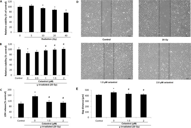
Cell viability, cytotoxicity and migratory ability of HUVECs following exposure to γ irradiation of different doses and celastrol treatment at different concentrations. (A) HUVECs were exposed to γ irradiation of 5 to 40 Gy. Cell viability was evaluated by MTT after 24 h. (B) HUVECs were exposed to 20-Gy γ irradiation, followed by treatment with celastrol at various concentrations (0, 0.5, 1, 1.5 and 2 µM). Cell viability was evaluated by MTT after 24 h. (C) Cells were exposed to 20-Gy γ irradiation, followed by treatment with celastrol at 1.5 and 2 µM. Cytotoxicity was determined by LDH release. HUVECs without celastrol treatment and γ radiation served as controls. Cell viability and cytotoxicity are expressed as a percentage of the control. (D) Images of HUVECs in the cell migration assay at 6 h after the removal of the inserts. The dishes were monitored under an inverted microscope (magnification, ×40). (E) Statistical results of the cell migration assay. The gap distances at 6 h after the removal of insert were used for comparison. All data are expressed as the mean ± SEM. ANOVA was used to determine statistical significance between groups followed by Tukey's post-hoc test. *P<0.05 vs. control; #P<0.05 vs. 20 Gy without celastrol treatment group. HUVECs, human umbilical vein endothelial cells; MTT, 3-(4,5-dimethylthiazol-2-yl)-2,5-diphenyltetrazolium bromide; LDH, Lactate Dehydrogenase; ANOVA, one-way analysis of variance.

