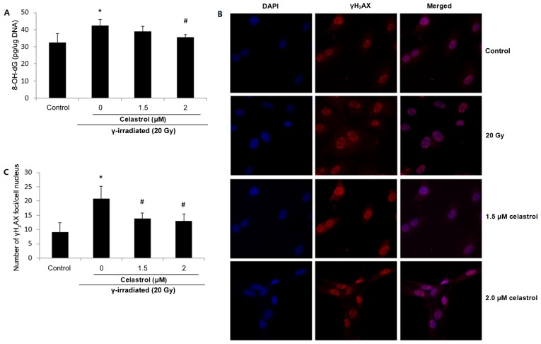
Figure 4.
Effects of celastrol on DNA oxidative damage in HUVECs. Cells were exposed to 20-Gy γ irradiation, followed by treatment with 1.5 µM celastrol. HUVECs without celastrol treatment and γ radiation served as the control. (A) Oxidative DNA damage was evaluated using an 8-OH-dG EIA kit. The absorbance at a wavelength of 420 nm was recorded and used to calculate the 8-OH-dG concentration based upon the standard curve. (B) Representative images of γH2AX fluorescence staining. DNA double-strand breaks were stained using anti-γH2AX antibody, and the nuclei were stained with DAPI. The images were examined under a fluorescence microscope (magnification, ×400). (C) The number of γH2AX foci was counted and compared. All data are expressed as the mean ± SEM. ANOVA was used to determine statistical significance between groups followed by Tukey's post-hoc test. *P<0.05 vs. control; #P<0.05 vs. 20 Gy without celastrol treatment group. HUVECs, human umbilical vein endothelial cells; 8-OH-dG, 8-hydroxy-2-deoxy Guanosine; ANOVA, one-way analysis of variance.

