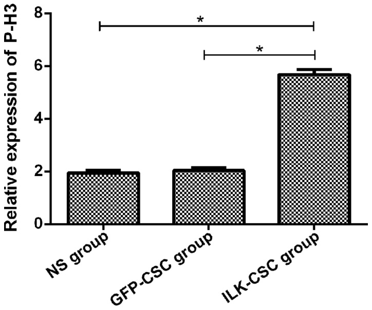
Figure 4.

The expression of P-H3 protein in the myocardial tissues of rats in each group. Results of immunohistochemical staining manifest that the expression level of P-H3 protein (5.68±0.19) in the myocardial tissues of rats in the ILK-CSC group is higher than that in the GFP-CSC (2.03±0.11) and NS (1.94±0.09) groups, and differences are statistically significant (*P<0.05). ILK, integrin-linked kinase; CSC, cardiac stem cell; GFP, green fluorescent protein; NS, normal saline.
