
Figure 4.
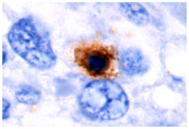
Representative immunohistochemistry image of latency-associated peptide+CD4+ T cells in HBV-infected hepatic tissues from the control group (high-power field; magnification, ×1,000). Positive cells were indicated by brown staining and blue staining indicates hepatic or tumor cells that are counterstained with hematoxylin in IHC images.