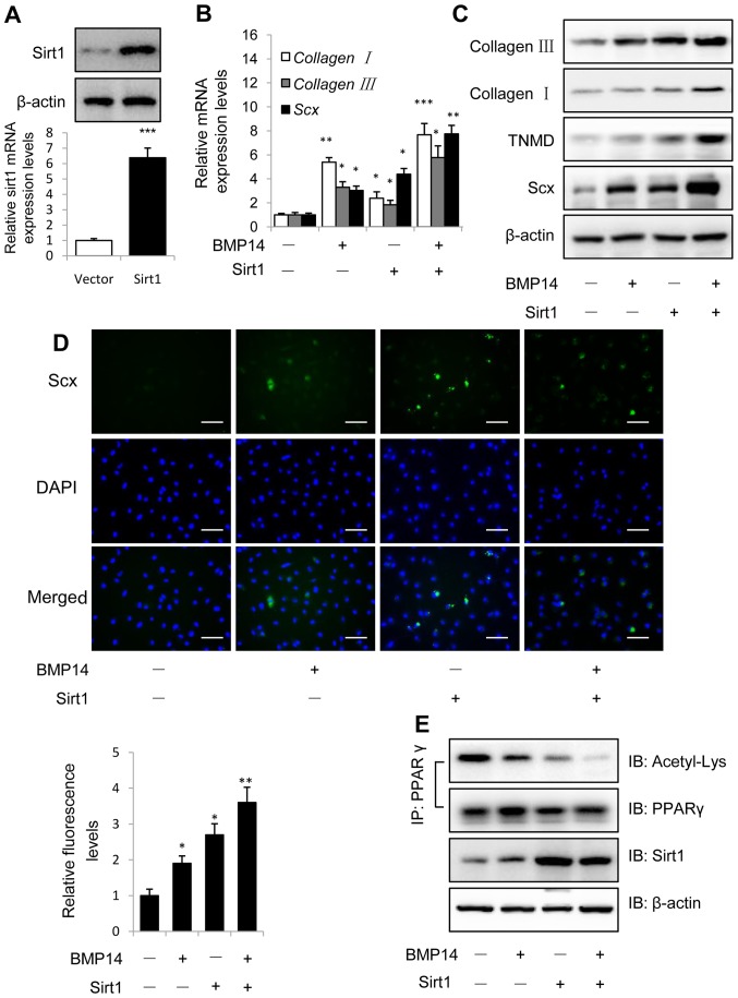
Figure 3.
Overexpression of Sirt1 promotes BMP14 induced tenogenic differentiation of BMSCs. (A) Sirt1 protein and mRNA levels were determined by western blot and RT-qPCR at 48 h post infection with the vector control or Sirt1 lentivirus. β-actin was used as the loading control. (B) mRNA expression of collagen I, collagen III and Scx and (C) protein expression of collagen I, collagen III, TNMD and Scx were detected using RT-qPCR and western blot analysis with or without 50 ng/ml BMP14 treatment for 48 h. (D) Fixed BMSCs were stained with Scx antibodies and Alexa Fluor 488 goat anti-rabbit secondary antibodies (green) and DAPI (blue). Scale bars, 200 µm. (E) PPARγ acetylation levels in BMSCs were analyzed using immunoprecipitation with anti-PPARγ antibodies and immunoblotting with anti-Acetyl-Lys and anti-PPARγ antibodies. The total cell lysate was immunoblotted with anti-sirt1 and anti-β-actin antibodies. Each bar represents the mean ± standard error of the mean. The results were repeated in three independent experiments. *P<0.05, **P<0.01 and ***P<0.001 vs. the control. RT-qPCR, reverse transcription-quantitative polymerase chain reaction; BMP, bone morphogenetic protein; BMSC, bone marrow mesenchymal stem cell; Sirt1, sirtuin 1; Scx, scleraxis; TNMD, tenomodulin; PPARγ, peroxisome proliferator-activated receptor γ; IP, immunoprecipitation; IB, immunoblotting.

